The famous brain map is now drawn to include complex movements
The organization of the primary motor cortex is not always so different for newborns and toddlers, but a new homunculus can help explain how targets work and how to control their movements
Sirigu said they thought they had all the information about the motor cortex. “But its organization is much more complex than we have traditionally thought.”
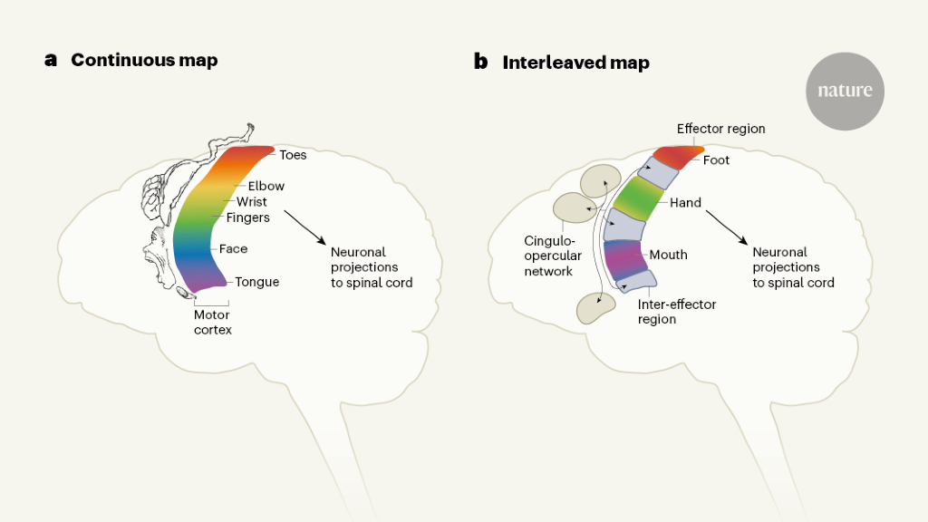
One of the most fundamental diagrams in neuroscience is thehomunculus. In countless textbooks, there is a picture of a constellation of body parts mapped onto a narrow strip of the brain and showing the same brain areas controlling each part.
A new theory was proposed based on data from above-elbow amputees. Theories suggest that there are two systems in the primary motor cortex, one for motor commands and the other for muscle synergies.
The researchers found the same regions in large data sets from previously scanned individuals. And when they scanned children, they found that a newborn hadn’t developed this brain network yet, whereas an 11-month-old and a 9-year-old had, supporting the theory that this network coordinates complex action plans, because newborns aren’t yet able to control their movements precisely, Dosenbach says.
So Dosenbach’s team was puzzled when they began seeing hints of a very different organization. The data from fMRI of individual brains was what led to the clues.
The findings could lead to changes in therapy for disorders of the primary motor cortex caused by stroke or injury. Fox says that a lot of the brain areas targeted for stimulation came from trial and error. A new homunculus could help explain how and why targets work.
What do epilepsy patients see in their brains? A study involving the motor cortex of Wilder Penfield, his son, and Nico Dosenbach
It must plan a trajectory, control dozens of muscles, make adjustments based on the feedback from eyes and fingers and maintain its focus on the goal: a great cup of coffee.
Blood pressure and heart rate can be influenced by the act of reaching for a cup of coffee. And the movement is seamlessly integrated into brain systems involved in planning, goals, and emotion.
According to the study, textbook depictions of a motor cortex with an “Intersection that is not going to be connected to a region that inquires ‘what am I going to do today'” is still accurate.
Evan Gordon, the study’s first author and an assistant professor of Radiology, says that the data from the scans leaves little doubt that there is a system. It was there even though we did not see it because of the things we’d learned in the first neuroscience class.
The chair of neurobiology at the University of Pittsburgh thinks that this is a really fundamental change in how he sees the motor cortex.
The finding involves a strip of brain tissue called the primary motor cortex. The main source of signals that control voluntary movements can be found in this area.
That view dates back to the 1930s, when Canadian neurosurgeon Wilder Penfield began mapping the brains of his epilepsy patients by applying electrical currents to areas in the motor cortex. Ultimately, Penfield identified segments that would reliably cause a foot, finger, or the tongue to move.
Gordon noticed that the MRI data suggested there were important areas between Penfield’s sections. These areas of cortex had lots of connections, but not to muscles. Instead, the connections led to areas all over the brain, including those that control internal organs like the heart and lungs.
At first, Gordon doubted what he was seeing. He wondered if it was odd that the data we have collected was in other people.
But if these segments of brain tissue weren’t for controlling muscles, what were they doing? To find out, the team turned to their lead scientist: Nico Dosenbach.
They put Dosenbach through complicated tasks, such as rotating his left hand in one direction and his right foot in the opposite direction. These tasks required his brain to plan his movements before carrying them out.
The motor cortex regions we found were more active during the planning stage, which was the right thing to point us in the right direction.
“There’s two interleaved systems,” Dosenbach says. Below the fingers the team would find an area involved in the whole body action.
And once again, Gordon says, they found evidence that the ribbon of motor cortex contained alternating areas: one for fine control of a specific muscle, then another keeping track of the entire body.
A system that weaves together movement and mental states also could explain why our posture changes with our mood, or why exercise tends to make us feel better.
How you move can affect the way you feel. And how you feel is going to have an impact on how you move,” Strick says. My mother told me to stand up straight and I would feel better. Maybe that’s true.