The face of plastic surgery could be changed by newly identified cells
Iridescent pearls? A cell biologist’s view of connective tissues implanted in human noses and adiposes
Maksim Plikus, a cell biologists at the University of California, Irvine and his colleagues examined the fat-filled cells under a microscope in order to understand their function. The team found that these cells have enormous intracellular structures called vacuoles that are filled with lipids. As a result, under the microscope, the cells “look like iridescent pearls”, Plikus says.
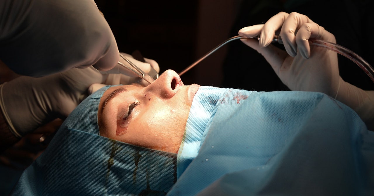
Cartilage transplants are a central part of many procedures, being used to fix cleft lips, correct missing ears, or to repair damage caused by cancer. They are often used in nose augmentations.
But the results aren’t always stellar. Surgeons often resort to transferring cartilage from the rib, which is stiff, or using silicone implants, with neither material matching the real thing. Implanted tissues are not flexible in the same way as those they’re implanted into, and don’t become part of the natively occurring tissue. “They often do not integrate, and they move around,” says Maksim Plikus, a cell biologist at UC Irvine. The nose needs a revision.
His team subjected the cells to a range of high techbiological profiles similar to personality tests. They proved that this bouncy connective tissue is neither typical chondrocyte nor adipocyte, according to results published today in Science.
The nose and ears are stretchy and squishy thanks to bubble wrap cells that provide extra cushion and structural support, according to a wide-ranging study.
“The idea that you regulate stiffness of tissue by controlling what’s inside the cell rather than through the extracellular matrix is new and interesting,” says Paul Janmey, a biophysicist at the University of Pennsylvania Perelman School of Medicine in Philadelphia. “That’s not typically the case, even in fat tissue.”
