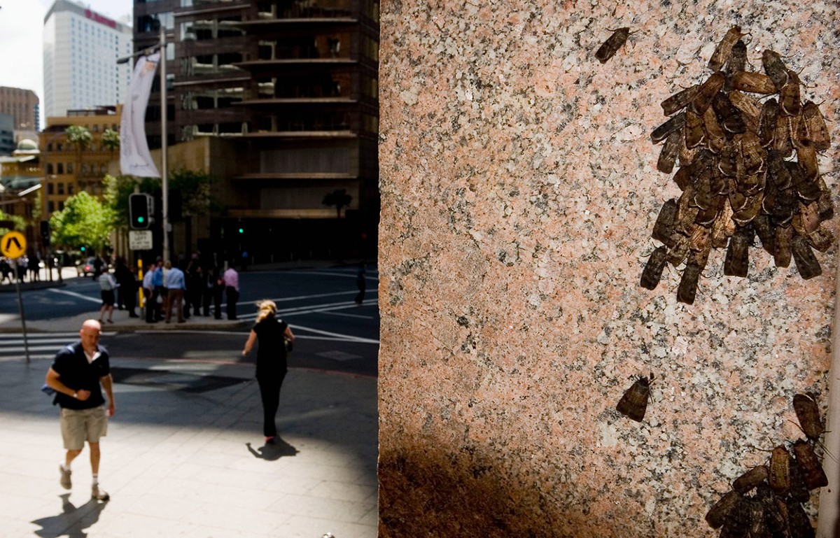
A compass is used by bhuong moths for long-distance navigation
Indoor behavioural and electrical experiments on dark-adapted moths in the early stages of radio-frequency disturbances at Glenhare, Adaminaby
A ferromagnetic-free laboratory located at Glenhare, Adaminaby was designed and built to house indoor behavioural and electrical experiments. 1c. The ground beneath the concrete slab contains a 6-mm thick, 30-mm wide and 12-m long copper strap that separated the main earth frombehaviour and electrophysiology. Background levels of radio-frequency disturbances at this rural site are extraordinarily low4. All of the experiments were performed on dark-adapted moths in darkness at night (beginning at least 1 h after sunset). Darkness was achieved with black-out blinds (to remove residual starlight and moonlight from outside) and dark cloth around the experimental apparatus (to shield from the minimal stray light emitted from the equipment).
The researchers used the magnetic field in the simulator to see if they could navigate without this cue. They also recorded the activity of visual neurons in the moths’ brains to investigate whether they have an internal representation of the starry sky.
Independent experiments were conducted under natural clear sky with new creatures used in each night. The data was obtained during a single night. Behavioural data under projected starry skies generated using Stellarium (Fig. 3c–f) were obtained in independent experiments run over several nights with each night’s cohort of moths used only once. These experiments were repeated with the same results over two separate spring and autumn seasons (2018 and 2019). All of the experiments deal with biological replicates. The samples were based on the availability of moths, with at least 40 being used in each experiment. Experiments were conducted without a specific randomization protocol. The images from the recordings and dye injections came from single visual cells in the brain, which were exposed to a projected starry sky. It was not possible to replicate each recording as it was a unique observation. This is due to the stochastic nature of intracellular recordings—single neurons were penetrated randomly from target brain regions that contain many thousands of cells.
The behavioural analysis used in this study has been previously described4,5. The instantaneous heading directions of a tethered flying mammal were recorded and saved in a text file, thanks to the software known asUSB1 Digital Explorer. The mean orientation of the tested moths was calculated using a mean orientation of each virtual flight path. Each plot contains both the mean and the r value for the flight path Vector of the moths. The flight trajectory of the moths have a mean direction as well as a mean directedness that we can take advantage of in our data. The R* value encodes the directedness of a population of tested moths and reveals the likelihood that the combined flight direction of these moths—each with its own direction and directedness—differs significantly from random.
The online data is used for the analyses. As there were no statistical differences in results obtained from male and female moths, the results were pooled.
Detection and incubation of reconstructed brains in the Bogong moth standard and using a Leica SP8 microscope
Brain samples were scanned with the 633 nm laser of a Leica SP8 confocal microscope and viewed with a ×20 oil-immersion objective (Leica Microsystems). For optimal resolution, the scan settings were set to 1,024 × 1,024 pixels, 12-bit pixel depth, 3 times line accumulation and 400 lines per s in the photon-counting mode of the hybrid detector. Neurons and relevant neuropils were then reconstructed in Amira v.5.3 (Thermo Fisher Scientific) and registered into the Bogong moth standard brain22,23.
After recording the cell, a positive current was applied to the implant to inject Neurobiotin. The electrode was then removed and the brain was dissected out of the head capsule. Brains were fixed in 4 °C overnight in paraformaldehyde solution (4% PFA in phosphate buffer) and then washed in 0.1 M PBS (4 times for 15 min). The retinas were removed during the washing. Brains were then incubated with streptavidin–Cy5 (Jackson Immuno Research, 1:1,000 in PBS with 0.3% Triton X-100) at 4 °C for 3 days and kept in the dark from this point onwards. After incubation, the brains were washed in PBS-Triton X-100 (6 times for 20 min) and PBS (2 times for 20 min) and then dehydrated in an increasing ethanol series (50%, 70%, 90%, 95% and twice at 100%). Brains were then transferred to a fresh mixture of methylsalicylate and ethanol (1:1) and, after 15 min, were left to clear in 100% methylsalicylate for 75 min. The cleared brains were mounted in Permount (Thermo Fisher Scientific) mounting media between two coverslips and left to dry for at least 2 days.
To see whether the eyes of the central brain could be seen from different parts of the Milky Way, we stimulated cells with signals from the stars and a projection screen above them. These cues mimicked aspects of the autumn Milky Way: its brightest region around the Carina nebula (revolving dot) and its stripe-like shape (rotating bar). The bar length and width was chosen to imitate the main stripe of our natural Milky Way. The mean grey level was adjusted so the artificial stimuli mimicked the stars and dark background sky in the natural environment, and that the intensities were adjusted to give a good mimic of them.
To maximize the success rate of the demanding intracellular recordings during the short migratory season, these experiments were performed both in the afternoon and during the natural nocturnal flight time of the moths. For afternoon experiments, we removed the two 1.2 log unit ND filters in front of the projector lens (as described above) to generate a starry sky projection around 250 times brighter than the one used at night (to account for the circadian-rhythm-induced light-adapted state of the moths). For night experiments, the ND filters were reinserted. No obvious differences were found in results obtained in the two situations. The moths were mounted onto the 3D-printed animal holder. A piece of the skin was taken from the head capsule to expose the brain, while the antennae were fixed with wax and a drop of wax. The neural sheath was digested with Pronase (Sigma-Aldrich) for about 30 s and then carefully washed. It was then removed using a pair of fine forceps. A second small hole was cut into the cuticle above the proboscis muscle and a chlorinated silver wire was inserted into this muscle to serve as reference electrode.
Most behavioural procedures used in this study have been described before. Before attachment of tethering stalks, moths were chilled in a freezer for 5–10 min to immobilize them. The scales on the moth’s dorsal thorax were removed by suction using a micro-vacuum pump (custom built by B.F.). A small circular footplate, which is ferromagnetic free and fashioned at its end, was fastened to the dorsal thorax using contact cement and a weighted-down plastic mesh. The day of the attachment of the stalks, the moths were tested.
Source: Bogong moths use a stellar compass for long-distance navigation at night
Circular statistical analysis of randomized stimulation using a custom-made UV LED-ring in front of a large projector for low-luminosity telescopes
For the fall and spring of 2018, the positions of the individual stars in the night sky image were redistributed randomly to new positions, and the resulting randomized images were saved as.PNG files. 9a). These featureless stellar conditions provided an identical stimulation intensity but provided no celestial spatial information. The randomized skies were used during spring and autumn so that they retained the stars, but also removed spatial variations in the night sky that could be used for orientation. Here, the stimulus image (of autumn 2019) was subdivided into squares with a size of 13 × 13 pixels, as the brightest star in the image had roughly these dimensions. The positions of these squares were randomly rearranged and a picture was created. During the spring of 2020 a final improved randomized stimulation was generated from the test using randomizing the visible stars. This was achieved by first detecting the position and size of each star in the test stimulus using a multiscale Laplacian of a Gaussian convolution of a greyscale version of the test stimulus, followed by local maxima detection. The resulting spatial information was then used to extract and save the image of each star from the natural night sky stimulus, before replacing them on a new background image, with a uniform colour and intensity equal to that of the mean of all pixels in the test stimulus that were not part of a star. This way, the location of the stars on the randomized image could be drawn from any desired distribution. In this case, a uniform distribution was used for the location of all but the brightest star, which was placed in the centre of the image.
As the projectors did not emit UV light, we installed a custom-made LED-ring (built by T. McIntyre; diameter, 120 mm; inner diameter, 50 mm) featuring eight UV LEDs (LED370E Ultra Bright Deep Violet LED, Thorlabs) in front of the projector. The UV intensity was brought into a quasi-natural range using a behavioural rig and several layers of filters that were fixed in front of the LEDs.
The data was analysed using the custom- written code in the MATLAB program. The maximum-likelihood estimation package is used for the circular statistical analysis. unimodal (models M2A, M2B, M2C) or bimodal (models M4A or M4B) were used to classify responses. Only responses that were classified as unimodal (models M2A, M2B or M2C in the R-library CircMLE, based on the Akaike information criterion) were analysed further with respect to the half-width of the rotation tuning curve, the SNR and the variability of the response.
“The first time we saw them flying under the night sky with no other cue and flying in the right direction we had to hold on to the edge of the table,” says study co-author Eric Warrant, an entomologist at Lund University in Sweden.
When the moths did not have access to the projection of the sky or to the electromagnetic field, they were unable to navigate at all. But with access to only the visual cue of the stars, they were able to navigate in the seasonally appropriate direction. They were also able to fly in the correct direction in the presence of an electromagnetic field, but no visible stars.

