There is a link between dad’s gut microbes and baby’s growth
Effects of gut microbiome on the placenta and placental development in prospective mothers: a two-sided Benjamini-Hochberg analysis
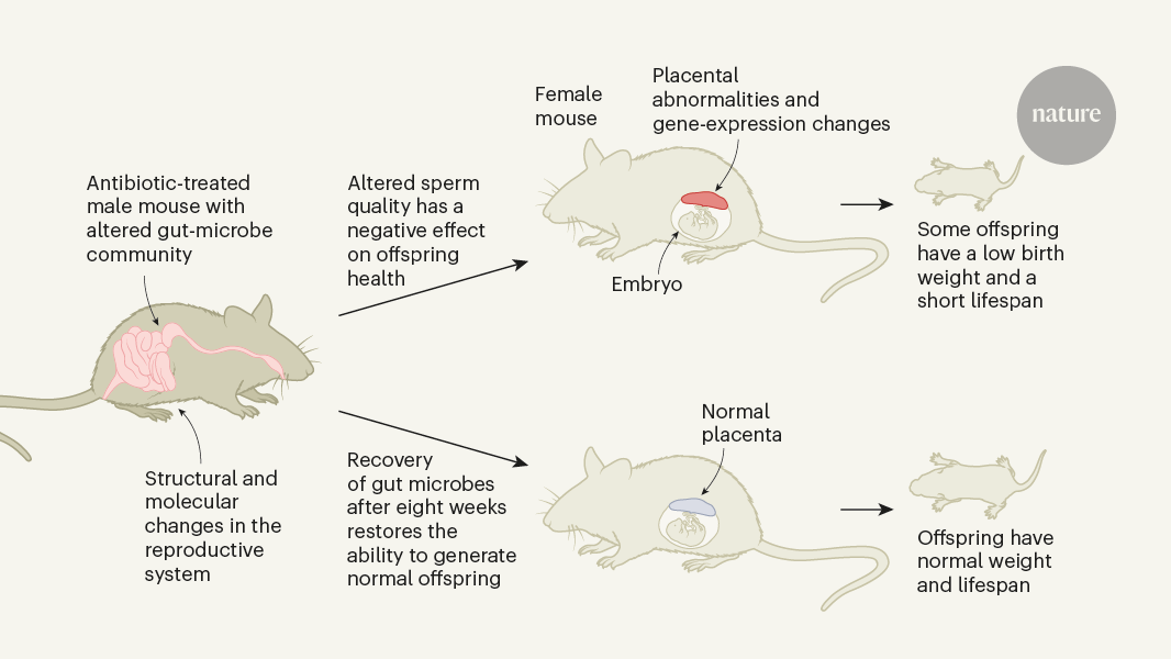
After changing the community of gut microbes in prospective fathers by administering either gut-specific (primarily non-absorbable) antibiotics or laxatives, the authors showed that the sperm from a father with a perturbed gut microbiome triggered changes in the placenta (which forms from cells of the embryo) that developed in its mating partner. The offspring of fathers with normal Microbiome had a higher chance of premature death than the offspring of fathers who had a lower Microbiome.
Crucially, the effect was reversed when the paternal gut microbiome was restored to normal by ceasing antibiotic exposure, and was recapitulated through in vitro fertilization using sperm from males harbouring the perturbed gut microbiome. Furthermore, the altered paternal microbiome was associated with changes in male reproductive tissue (smaller testes and seminiferous tubules with a swollen (vacuolated) appearance and thinner than normal layers of epithelial cells). The authors observed intact genomic parental-specific expression of genes (imprints) but altered transcriptome, metabolome and methylome profiles (relating, respectively, to gene expression, production of metabolite molecules and the attachment of methyl groups to DNA); these profiles were of unknown relevance to the observed outcome.
The treated and control group had their analysis calculated using a Fisher’s exact test. All P values were FDR-corrected for several hypotheses testing using the Benjamini–Hochberg procedure (stats::p.adjust function in R with BH parameter).
Statistical significance of the feature intensity differences was assessed using a two-sided t-test (stats::t.test function in R) of log-scaled data and P values were FDR-corrected for several hypotheses testing using the Benjamini–Hochberg procedure (stats::p.adjust function in R with BH parameter). Adjusted P values and foldchanges were visualized using Volcano plots. Mass, retention time, HMDB annotation, class, superclass and composite spectrum were retrieved for all features showing significantly different intensity between the treated and the control group and an absolute foldchange greater than 2.
Annotation was obtained from the Human Metabolome Database (HMDB), where it could be retrieved by searching for the exact same mass of a metabolite and falling within a window from the mono-isotopic mass. When present, several annotations were retrieved. Only features being annotated were retained for the downstream analysis. Moreover, for each feature, metabolite class and superclass were retrieved, when present, from the HMDB.
Alignment reads were trimmed using Trim Galore (v.0.6.3)79, then the first ten 5′ bases of both reads removed with Cutadapt (v.2.3)80. Reads were aligned to M. musculus refence genome (mm10) using bwa mem (BWA-0.7.17)81 before filtering with samtools (v.1.10)82 view with the flags ‘-h -F 256 -f 2 -q 30’ and deduplicated with Picard toolkit (v.2.9.0) MarkDuplicates83. SNPs and small INDELs were called using GATK (v.4.1.6.0)84 HaplotypeCaller. The QUAL of people with more than one individual was taken into account, and those with less than 0.2 or low site coverage were also removed. Variant functional region was annotated using ANNOVAR (2020Jne07) annotate_variation.pl85. The structure was called with the Delly2 call.
The last two bp of a single-end read were thrown out, as well as the first 8bp from the 5th end and the first 8bp from the 5′s end, in order to match the length of the fastq sequence. Cytosine methylation status was extracted from mapped reads using the Bismark methylation extractor tool. Five biological independent replicate datasets for each condition were used to analyse genome-wide methylation calls. To identify DMRs, the genome was first binned into sliding tiles containing 50 consecutive CpGs and their methylation status determined using the DNA methylation pipeline. The minimum of 10 reads is required for read-depth sensitive logistic regression to be effective for identified DMRs. Seqmonk calculated the level at specific features using the DNAMethylPipeline.
To investigate significant differences, data were processed using DESeq2 and the negative binomial test used to identify significant differences after Benjamani–Hochberg multiple test correction.
Deuteron-Antiproton Genomics (DEGs) on Mouse Embedded in the Musculus RNA Database using the FindMarkers Function
Adaptors were removed using fastx-clipper; fastq files were converted to fasta using a custom perl script and reads of 18–33 nucleotides in length were retained using a custom perl script. The best alignment of the reads required 0 mismatches and was reported by bowtie. The files were converted using bedtool v.2 and samtools v.1.9. To quantitate the number of reads per overlap of the known M musculus miRNAs, intersectBed -c was used. The tRNAs were obtained by using intersectBed -c to count the number of reads overlapping a bed file documenting predicted mouse tRNA coordinates downloaded from the tRNA scan database (http://gtrnadb.ucsc.edu/genomes/eukaryota/Mmusc10/). Ref’s piwi-Interacted RNA coordinates were taken. 78 Liftover converted the letter to the letter. The piRNAs were quantitated by selecting RNAs between 26 and 32 nucleotides long with a U as the first nucleotide and intersecting these RNAs with the piRNA coordinates using intersectBed -c.
All expressed genes were used in the principal component analyses of transcriptomes. These were defined by having an average of at least two replicates in all the samples.
DEGs were determined using the DESeq2 package (v.1.24.0), inputting raw mapping counts and applying a multiple-testing adjusted P value (false discovery rate (FDR)) < 0.05 significance threshold. An extra foldchange filter of more than 2 was applied to generate final DEGs.
For each cell type identified in the dataset, the FindMarkers function was used to identify DEGs between nABX and CON cells using logfc.threshold = 0.25 and p_val_adj < 0.01.
The dataset was annotated using uniquely expressed marker genes. The clusters showing no uniquely expressed genes were ignored. The germ and somatic cells were split into different objects to make more fine-grained annotations.
The nABX and CON samples were then integrated using the canonical correlation analysis approach at default parameter settings as recommended by the Seurat package.
To assess the effect on the body weights of sires treated with nABX, the weights of CON sires were compared to those of nABX sires. The testes were carefully collected from CON and nABX treated mice to assess the impact on reproductive organs.
As described previously54, this method of F1 offspring generation follows five critical steps: (1) superovulation of oocyte-donor females; (2) preparation of fresh sperm from sire males; (3) collecting isogenic oocyte from donor females; (4) in vitro sperm–oocyte fertilization; (5) embryo transfer to pseudopregnant CD1 or C57BL/6J surrogate mothers. This process was done.
To separate the potential indirect impact on offspring of females with a male who is dysbiotic we use a co strategy combined with control male mating. A male with or without previous nABX exposure was placed with a virgin female during the day, recapitulating full parental co-exposure. The males were removed from the cages in order to prevent them from having sex at night and females to maximize detection of coprophagic effects. Each day at 16:00, females were checked for vaginal status, all females with positive signs of mating (vaginal plugs) were excluded from the study, whereas all females that were vaginal plug negative were held in the male cages. In a 4 day cohousing period, mice were kept in standard optimal environments with access to food and water. After 4 days of cohousing, all plug-negative females (exposed to nABX or CON males) were mated with conventionally raised 11-week-old C57BL/6J male mice (independent naïve males) to determine whether any indirect effects of previous nABX male exposure could be detected in F1 phenotypes.
Sperm–oocyte fertilization: from capacitated sperm in TYH-MBCD drops, a 10 μl of sperm suspension was taken from the peripheral part of the pre-incubation drops which contain the most motile sperm. Aliquoted sperm was then transferred from CON mice into the center of the fertilization dish containing oocytes. The sperm–oocytes fertilization dishes were cultured at 37 C under 5 CO2 for a longer period of time. After 3–4 h of incubation, the presumptive zygotes were washed three to four times with 150 μl drops of M2 medium and cultured overnight in four-well NUNC plates with 500 μl of KSOM medium supplemented with amino acids (KSOMaa) at 37 °C under 5% CO2 and 5% O2 incubator. The fertilized two-cell stage embryos were transferred to the serous uterus of the female surrogate mice.
The method for calculating sperm count was described by the ref. It was used for sperm counts. The spermsuspension was prepared by mincing cauda in 1 liter of PBS. The suspension was pipetted and filtered through 80 μm nylon mesh to remove tissue fragments. An aliquot (0.05 ml) from the sperm suspension (1 ml) was diluted with 1:40 PBS (pH 7.2) and mixed thoroughly. The sperm suspension was counted using Bürker chamber after discharging a few drops of the suspension. To calculate sperm count per mouse, the total number of sperm in each square, was averaged and compounded by the dilution factor 104 to arrive at a figure.
There were slight alterations to the description of the seminal vesicles collected in the flow-hood. One experimental animal was put in the hood for each sample collection. The experimenter wore sterile clothing, gloves, mask, and took all necessary precautions to make sure the experiment was free of parasites. In brief, the mice were placed in a laminar flow-hood that had been previously cleaned with 70% ethanol and 2% Virkon followed by 2 h of ultraviolet radiation. To collect seminal vesicles aseptically, mice were euthanized by cervical dislocation and their abdominal areas were cleaned with 70% ethanol and incised with sterilized tweezers and scissors. After the seminal vesicles were excised, they were placed in a sterile 2 liter Eppendorf tube which was frozen for 80 C. Note, dissection tools used in this sample collection were used only once per mouse.
Blood samples were collected from saphenous vein microvette blood collection tubes (Microvette 100K3E SARSTEDT, catalogue no. 20.1278.100) and centrifuged at 5,000 rpm for 10 min at 4 °C; plasma was obtained and stored at −80 °C until assayed.
Placenta samples were fixed in 4% paraformaldehyde at 4 C and then sectioned into 7-m thick slices. Sectioned samples were mounted on slides and dried at 40 C. The dried sections were subjected to heat-induced antigen retrieval using citrate buffer at pH 6.0, incubated overnight at 4 °C with anti-mouse VE-cadherin (Thermo Fisher Scientific; eBiosciences. catalogues no.14-1441-81, 2.5 g ml1) and anti-Rat-AlexaFlu 568 have been detected. A-11077; 4 μg ml−1). There were four compounds that were counterstained with, 4′,6 diamidino-2-phenylindole (DAPI). D1306; 5 μg ml−1). Slides were washed before being mounted with ProLong Glass (Thermo Fisher Scientific; catalogue no. P36983) and imaged on a Leica THUNDER Imager Live Cell with a ×20 objective (numerical aperture = 0.8). The areas of labyrinth zone and whole placenta were measured with the help of image analysis software.
The concentration of a specific targetProtein in biological samples can be determined by using ELISA kits. Frozen plasma and testis samples were used for the measurement of leptin (Lep) in F0 sire males, whereas F1 placenta tissues were used for the measurement of placental growth factor (Plgf) and soluble fms-like tyrosine kinase-1 (sFlt1). Mouse leptin kit has mouse. A person is named ELISA. Following the manufacturer’s instruction, both the Kit MP200 and mouse were used for quantification of Lp, plgf, and sFlt1. The samples, standards and control were all prepared using the kits and then tested in duplicate with a number of biological replicas. The data was put to use by the regression model to find the target concentrations.
Source: Paternal microbiome perturbations impact offspring fitness
Measurement of antibiotic residues in distal tissues of F0 sires with an Agilent 6550 iFunnel mass spectrometer
To screen for antibiotic residues and test whether nABX or its residues are detectable in distal tissues or the circulatory system we took independent and complementary approaches.
Samples for the quantification of antibiotics in testis tissues were prepared as described in the section on Metabolomics below. Extracted and dried samples were resuspended in 20 μl of methanol:water (1:1) and 10 μl of samples were directly injected into an Agilent 6550 iFunnel qToF mass spectrometer. The standard used to prepare the curves was 5.1– 9.1 pmol for bacteriocin, 7.5–112.5 pmol for primaricin, and 5.3–10.5 pmol for neomycin. Quantification of antibiotics was performed using the MassHunter Quantitative Analysis Software (Agilent Technologies, v.10.0) and limit of detection was determined by linear regression using R.
PremiTest kit (R-Biopharm) was used for the determination of nABX or avaABX residues in testis samples stored at −80 °C, which was performed in accordance with the manufacturer’s instructions for use in meat. The test is based on thermophilic bacterium strains which are sensitive to several antibiotics and other compounds. The testis were frozen in ice, washed in PBS and then homogenized using a tissue grinder containing 200 grams of 1. There were two ways of controlling the amount of water used: sterile water and nABX cocktail mixed with water and taken out of the water. The sample solution was pipetted onto the agar in the Ampoule, which was allowed to stand at room temperature for 20 minutes before prediffusion. After the prediffusion, the sample solution was flushed out of the ampoule by washing twice with water and covered with foil to avoid evaporation. The positive control on the test was turned from purple to yellow after a few hours in Eppendorf block heater. The results of the block heating were interpreted on the basis of the kit bicolour indicator after the Ampoules were removed.
Fresh faecal samples from each individually housed F0 sire males were collected at several different times before and after nABX administration, at the end of a 6 week treatment before setting up mating, and after nABX withdrawal. At the time of the F1 offspring’s birth, fecal samples from the dams were collected. To collect faecal samples, faeces were put into sterile cages with food and water for 2 h, then faecal pellets were collected with a sterilizer and stored in the freezer for 80 C.
The samples were stored at a temperature of 80 C until they were processed. Microbial DNA extraction from faecal specimens (about 200 mg from F0 parent and about 100 mg from F1 offspring) was performed using QIAamp PowerFecal Pro DNA Kit (QIAGEN) according to the manufacturer’s instructions. Briefly, faecal samples processed with the DNA extraction kit were added to a PowerBead beating tube and rapidly homogenized on a Vortex Adapter (QIAGEN catalogue no. 13000-V1-24) using Vortex-Genie 2 mixer (Scientific Industries). After cells are lysed and potential inhibitors removed, total genomicDNA is captured on a Silica membrane washed and eluted for downstream gut microbiota profiling.
The relative abundance of 16S rRNA genes was determined using a standard curve based on serial dilutions of the control sample and real-time qPCR.
Targeted amplification of the 16S rRNA V4 region (primer sequences F515 5′-GTGCCAGCMGCCGCGGTAA-3′ and R806 5′-GGACTACHVGGGTWTCTAAT-3′ (ref. The KAPA HiFi HotStartPCR mix (Roche) was used in the procedure with minor modifications to the manufacturer’s instructions. The European molecular Biology Laboratory in Heidelberg used size-selective magnetic beads and a Illumina MiSeq to sequence the products.
The raw rRNA reads were trimmed, denoised and cleansed before being put in a sealed container. 61). The resulting amplicon sequence variants were then clustered into operational taxonomic units (OTUs) at 98% sequence similarity using an open-reference approach: reads were first mapped to a preclustered reference set of full-length 16S rRNA sequences at 98% similarity using MAPseq62. Reads that did not confidently map were aligned to bacterial and archaeal secondary structure-aware rRNA models using Infernal63 and clustered into OTUs with 98% average linkage using hpc-clust64, as described previously65. The resulting OTU count tables were noise filtered by asserting that samples retained at least 1,000 reads and taxa were prevalent in at least two samples; these filters removed 58% of spurious OTUs but only 0.09% of total reads from the dataset.
Local sample diversities were calculated as OTU richness, exponential Shannon entropy and inverse Simpson index (corresponding to Hill diversities of order 0, 1 and 2 (ref. 66) as average values of 100 rarefaction iterations to 5,000 reads per sample. Community diversity was calculated by Bray–Curtis dissimilarity. Trends in community composition were quantified using ordination methods (principal coordinate analysis, distance-based redundancy analysis) and tested using permutational multivariate analysis of variance (PERMANOVA67, as implemented in the R package vegan68.
Source: Paternal microbiome perturbations impact offspring fitness
Amplification of microRNA isolated from sperm samples by the QIAGEN Hilden kit in conjunction with TaqMan Nanoscale Master Mixing
Independent males from the small RNA-seq samples were used to extract purified sperm total RNA with the miRVana microRNA isolation kit, applying minor modifications to ensure complete lysis. Quantification of miRNA and tRNA was performed using predesigned (miR-141-3p) and custom-designed (tRNA-Gly-GCC-2) According to the manufacturer’s protocol, seven ng of cDNA from CON and nABX were used to create the TaqMan assays. The quantitative PCR with reverse transcription was performed in 15 μl reactions using TaqMan Fast Advanced Master Mix, following the standard programme (10 min at 95 °C; 15 s at 95 °C; 1 min at 60 °C, for 40 cycles). snoRNA 202 was used as a source of control for the miRNA and tRNA levels. Relative quantities were normalized to endogenous control values and foldchange calculated by means of the 2−∆∆Ct method. Amplification efficiency of TaqMan probes was confirmed by serial dilution of template runs.
Testicular single-cells resuspended in PBS + 0.04% BSA were adjusted at a concentration of about 1,100 cells μl−1 for loading on the 10x Chromium chip. Cell capturing and library preparation was carried out according to instructions in the kit. There is a user guide for the dual index. After a sample was collected, about 6,000 cells were targeted and then used for library amplification. The resultant libraries were size selected, pooled and sequenced using Illumina NextSeq 2000 P2 flowcell (100CYC) was used for the protocol.
The QIAGEN Hilden is a purification kit for sperm genomic DNA which was used to extract total sperm DNA. The DNA extraction was performed as follows. Frozen pure sperm samples stored at −80 °C were thawed in ice; 100 μl of DNA extraction buffer (20 mM Tris Cl is 20 mM. EDTA, 200 mM NaCl, 4% SDS) containing 80 mM dithiothreitol and 12 μl ml−1 of proteinase K was added to the sample; and then incubated at 56 °C on Eppendorf thermomixer until complete sperm lysis was assured (about 1 h), shaking occasionally during incubation to disperse the sample. After incubation, the user-developed protocol DY03 (QIAGEN) for purification of total DNA from animal sperm was applied. BS-Seq libraries were constructed according to the manufacturer’s instructions using TruSeq DNA Methylation Library Prep Kit (Illumina). Multiplexing libraries were performed on an Illumina NextSeq 500.
Following the manufacturer’s instructions, the gDNA was taken from the offspring of control or nABX-derived F1 in the normal or SGR way. The quantity and quality of DNA was evaluated on a NanoDrop Spectrophotometer and Agilent TapeStation system (Agilent) to ensure DNA integrity number > 7. The library Prep kit was followed by all manufacturer’s guidelines. Amplified libraries were multiplexed and sequenced on an Illumina Hiseq 4000 (PE150) to appropriate depth.
Source: Paternal microbiome perturbations impact offspring fitness
Mass calibration for LC–MS using the Mass Hunter Qualitative Analysis Software: absolute height determination, peak alignment and alignment determination using Mass Profiler Professional
Fisher Scientific bought all the chemicals for LC–MS analysis. Standards for online mass calibration were purchased from Agilent Technologies.
The MassHunter Qualitative Analysis Software was used to extract data from LC–MS. The settings were applied to the absolute height: 5,000 counts, limit assigned charge states to 1, and only H+ charged molecule included with compound quality scores greater than 80%. Peak alignment and identification were carried out using Mass Profiler Professional (Agilent, v.15.1) with default parameters: mass tolerance of 2 mDa or 20 ppm and retention time tolerance of 0.2 min or 2%. Extracted and aligned features were exported as .csv file for further data analysis
Source: Paternal microbiome perturbations impact offspring fitness
Multiplicity analysis and data analysis using Graphpad Prism and R for the study of paternal microbiome perturbations impact offspring fitness
Statistical analyses was performed using Graphpad Prism v.8.4.3 graphical software and in R v.3.6.2. The replicate number is dependent on the number of fathers that are uniquely exposed and not the number of offspring, which is greater. We typically generate offspring n > 150 per experiment in order to capture effect size and partially penetrant phenotypes but each offspring is Nested into a single N value. A nested t-test compares the means of two groups, one control and one dysbiotic, for which there are a combination of factors. This is necessary as using individual offspring as independent variables in intergenerational studies will lead to inflated alpha error rates and spurious significance. The labyrinth zone and fetoplacental ratio were analysed by twotailed unpaired t-test. The Baptista–Pike method was used to calculate the odds of the ORs and the chi-squared test was used to determine the CIs. The log-rank test was used to generate survival analysis curves. ELISA calibration curves were interpolated with a Hyperbola (X is concentration) nonlinear regression model fit (R2 > 0.99 was acceptable curve fit).
Raw reads were aligned and mapped using the count module in 10x Genomics Cellranger 6.1.2 (ref. 75) to the mm10 transcriptome assembly (2020-A) with default parameters.
The Seurat package was used to perform all the subsequent steps. The number of unique genes detected, the percent of Mito and ribosomal rate were the first parameters to be used in the screening of cells. The samples were grouped using the default clustering approach. In both circumstances, clusters with no uniquely expressed genes were discarded.
Source: Paternal microbiome perturbations impact offspring fitness
A study of testicular deficiency in CD1 and CD1 inbred mice for IVF: a laboratory animal management and ethics committee study
All experiments involving mice were carried out in accordance with the approved protocol and guidelines by the laboratory animal management and ethics committee of the European Molecular Biology Laboratory (EMBL) under license no. The Italian Ministry of Health is under authorization code no. 308/2021-PR. The inbred C57BL/6J strain was used as the primary mouse model, whereas CD1 IGS or C57BL/6J dams were used as surrogate mothers for IVF. There were C57BL/6J mice that were deficient in leptin used to study the effects of testicular leptin deficiency. Mice were housed under a 12 h light/dark cycle (from 07:00 to 19:00), with ad libitum access to regular diet and water. Standard chow contained 18.5% protein, 5.3% fat, 4% fibre and other nutritional additives in pellet form and is suitable for long-term maintenance, breeding, lactation and gestation periods (NFM18, Mucedola).